PDF] Brain Tumor Segmentation of MRI Images Using Processed Image Driven U-Net Architecture
Por um escritor misterioso
Last updated 26 outubro 2024
![PDF] Brain Tumor Segmentation of MRI Images Using Processed Image Driven U-Net Architecture](https://d3i71xaburhd42.cloudfront.net/c750894747d2b3f841de55922b2b68794295de27/7-Table3-1.png)
A fully automatic methodology to handle the task of segmentation of gliomas in pre-operative MRI scans is developed using a U-Net-based deep learning model that reached high-performance accuracy on the BraTS 2018 training, validation, as well as testing dataset. Brain tumor segmentation seeks to separate healthy tissue from tumorous regions. This is an essential step in diagnosis and treatment planning to maximize the likelihood of successful treatment. Magnetic resonance imaging (MRI) provides detailed information about brain tumor anatomy, making it an important tool for effective diagnosis which is requisite to replace the existing manual detection system where patients rely on the skills and expertise of a human. In order to solve this problem, a brain tumor segmentation & detection system is proposed where experiments are tested on the collected BraTS 2018 dataset. This dataset contains four different MRI modalities for each patient as T1, T2, T1Gd, and FLAIR, and as an outcome, a segmented image and ground truth of tumor segmentation, i.e., class label, is provided. A fully automatic methodology to handle the task of segmentation of gliomas in pre-operative MRI scans is developed using a U-Net-based deep learning model. The first step is to transform input image data, which is further processed through various techniques—subset division, narrow object region, category brain slicing, watershed algorithm, and feature scaling was done. All these steps are implied before entering data into the U-Net Deep learning model. The U-Net Deep learning model is used to perform pixel label segmentation on the segment tumor region. The algorithm reached high-performance accuracy on the BraTS 2018 training, validation, as well as testing dataset. The proposed model achieved a dice coefficient of 0.9815, 0.9844, 0.9804, and 0.9954 on the testing dataset for sets HGG-1, HGG-2, HGG-3, and LGG-1, respectively.
![PDF] Brain Tumor Segmentation of MRI Images Using Processed Image Driven U-Net Architecture](https://www.medrxiv.org/content/medrxiv/early/2022/11/04/2022.11.03.22281923/F1.large.jpg)
Comparing 3D, 2.5D, and 2D Approaches to Brain Image Segmentation
![PDF] Brain Tumor Segmentation of MRI Images Using Processed Image Driven U-Net Architecture](https://www.science.org/cms/10.1126/sciadv.add3607/asset/f006810b-5ff3-4034-a144-00b49132fbcb/assets/images/large/sciadv.add3607-f1.jpg)
SynthSR: A public AI tool to turn heterogeneous clinical brain scans into high-resolution T1-weighted images for 3D morphometry
![PDF] Brain Tumor Segmentation of MRI Images Using Processed Image Driven U-Net Architecture](https://d3i71xaburhd42.cloudfront.net/d12c02378276e1fb4f8dd00fd8b25e9761866a56/3-Figure1-1.png)
PDF] Attention Gate ResU-Net for Automatic MRI Brain Tumor Segmentation
![PDF] Brain Tumor Segmentation of MRI Images Using Processed Image Driven U-Net Architecture](https://d3i71xaburhd42.cloudfront.net/420f3f1078a6d8e0696572c032877079286051c6/3-Figure1-1.png)
PDF] Brain Tumor Segmentation Using Convolutional Neural Networks in MRI Images
![PDF] Brain Tumor Segmentation of MRI Images Using Processed Image Driven U-Net Architecture](https://pub.mdpi-res.com/computers/computers-10-00139/article_deploy/html/images/computers-10-00139-ag.png?1635497073)
Computers, Free Full-Text
![PDF] Brain Tumor Segmentation of MRI Images Using Processed Image Driven U-Net Architecture](https://content.iospress.com/media/xst/2020/28-1/xst-28-1-xst190552/xst-28-xst190552-g011.jpg)
Improving brain tumor segmentation on MRI based on the deep U-net and residual units - IOS Press
![PDF] Brain Tumor Segmentation of MRI Images Using Processed Image Driven U-Net Architecture](https://www.mathworks.com/help/images/segment3dbraintumorsusingdeeplearningexample_01_ja_JP.png)
3-D Brain Tumor Segmentation Using Deep Learning - MATLAB & Simulink Example
![PDF] Brain Tumor Segmentation of MRI Images Using Processed Image Driven U-Net Architecture](https://www.thelancet.com/cms/asset/da1b6d26-9d96-4e32-8a8b-0f4bc8ed2d85/gr1.jpg)
Deep-learning-based synthesis of post-contrast T1-weighted MRI for tumour response assessment in neuro-oncology: a multicentre, retrospective cohort study - The Lancet Digital Health
![PDF] Brain Tumor Segmentation of MRI Images Using Processed Image Driven U-Net Architecture](https://www.med.upenn.edu/cbica/assets/user-content/images/BraTS/brats-tumor-subregions.jpg)
3D MRI Brain tumor segmentation, U-NET
![PDF] Brain Tumor Segmentation of MRI Images Using Processed Image Driven U-Net Architecture](https://static.hindawi.com/articles/jhe/volume-2022/4189781/figures/4189781.fig.004.jpg)
U-Net-Based Medical Image Segmentation
Recomendado para você
-
 how to do brain test level 191|TikTok Search26 outubro 2024
 -
 Brain Test Level 191, 192, 193, 194, 195, 196, 197, 198, 199, 200 Answers26 outubro 2024
 -
Brain Out Level 191, 192, 193, 194, 195, 196, 197, 198, 199, 200 Solutions., Brain Out Level 191, 192, 193, 194, 195, 196, 197, 198, 199, 200 Solutions. : By BRAIN Game Solution26 outubro 2024
-
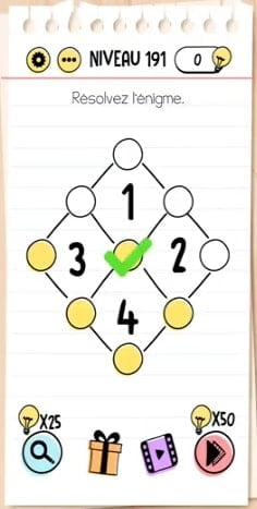 solution Brain Test Niveau 191 - android & iphone26 outubro 2024
 -
 Brain Test 4 Levels 191, 192, 193, 194, 195 Answers26 outubro 2024
 -
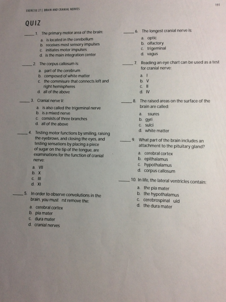 Solved 191 EXERCISE 27, BRAIN AND CRANIAL NERVES QUIZ 1.26 outubro 2024
 -
 Case 12: left visual'spatial neglect observed in the lime crossing test26 outubro 2024
 -
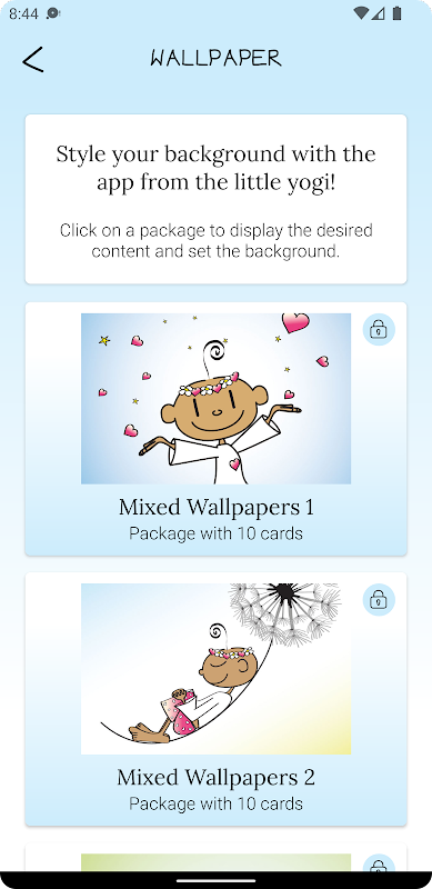 The Little Yogi - APK Download for Android26 outubro 2024
 -
 JOGO - QUEM É ELA? Coração de Educador26 outubro 2024
 -
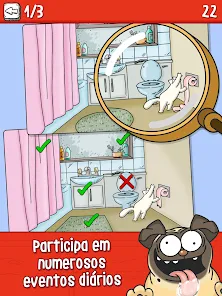
Simon's Cat - Crunch Time – Apps no Google Play26 outubro 2024

você pode gostar
-
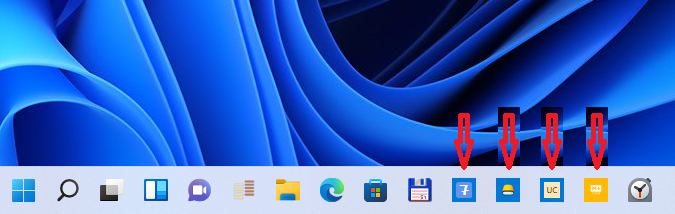 Tiny taskbar icons in Windows 11 - Microsoft Q&A26 outubro 2024
 -
 Nos pênaltis, Brasil perde para Croácia e está fora da Copa do Mundo - Jornal Mundo Lusíada26 outubro 2024
 -
 Tsukasa Mikogami (Choujin Koukousei-tachi wa Isekai demo Yoyuu de26 outubro 2024
 -
 Certificado de vacinação da filha de Bolsonaro foi gerado em inglês um dia antes de viagem aos EUA - Estadão26 outubro 2024
 -
Wotakoi: Love Is Hard For An Otaku OVA 3 on Vimeo26 outubro 2024
-
 Boruto: Naruto Next Generations, Wiki26 outubro 2024
 -
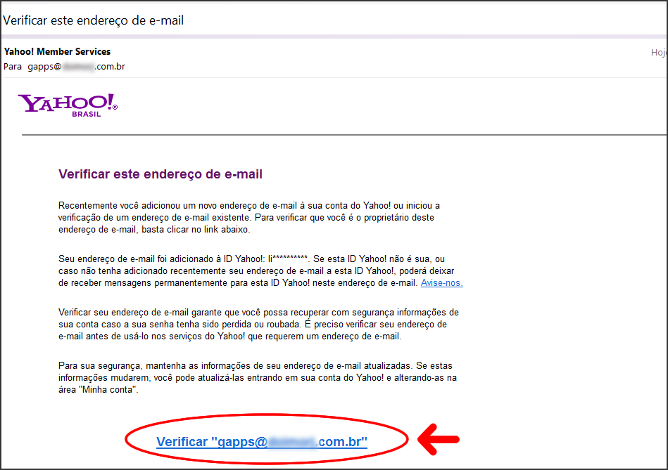 Como acessar minhas mensagens de e-mail pelo webmail do Yahoo! Mail? :: Ajuda Online Fastcommerce26 outubro 2024
 -
 Looking for Roblox Hungry Orca Code, can trade almost any other prime code : r/primegaming26 outubro 2024
 -
 Jurassic World Bite Fight Tyrannosaurus Rex GCT91 - ToysChoose26 outubro 2024
 -
 Frete grátis americano resina decoração estilo moda xadrez 3 peças por conjunto com rainha rei e cavalo em de…26 outubro 2024